You’ll often notice subtle mucocutaneous and systemic findings from iron deficiency—pallor, angular cheilosis, glossitis, koilonychia, hair thinning, fatigue, exertional dyspnea, tachycardia, cognitive slowing, pica, or restless legs—that correlate with depleted iron stores and reduced hemoglobin. Presentation varies by age and chronicity, so you’ll need targeted screening with CBC, ferritin, transferrin saturation, and CRP to guide management and further evaluation.
Common Physical Symptoms of Iron Deficiency

Although symptoms vary with severity and chronicity, iron deficiency typically produces a reproducible constellation of physical signs: progressive exertional dyspnea and generalized fatigue from impaired hemoglobin-mediated oxygen delivery; conjunctival and palmar pallor reflecting reduced hemoglobin and capillary oxygenation; tachycardia and exertional tachypnea as compensatory responses; koilonychia and brittle nails from impaired keratinization; glossitis, angular cheilosis, and atrophic mucosa indicating epithelial turnover deficits; and neurobehavioral manifestations such as pica and restless legs syndrome linked to altered neurotransmitter synthesis and iron-dependent enzyme activity. You should prioritize objective assessment—CBC, ferritin, transferrin saturation—and correlate with functional metrics like VO2. Examine mucocutaneous features and nails systematically. Initiate targeted replacement or investigate occult losses when biochemical indices confirm deficiency, and monitor hemoglobin plus ferritin kinetics to gauge therapeutic efficacy serially.
Less Obvious or Subtle Warning Signs
You may experience nocturnal periodic limb movements and an irresistible urge to move your legs (restless legs syndrome), a finding that is epidemiologically associated with iron-deficiency states. You might also develop pica, defined by compulsive consumption of nonnutritive substances (e.g., ice, clay, starch), which often correlates with low ferritin levels. Additionally, you can present with glossitis and angular stomatitis—tongue erythema, atrophy, and mucosal soreness—reflecting epithelial changes linked to iron depletion.
Restless Legs at Night
When iron stores fall below physiologic requirements, dopaminergic neurotransmission in the central nervous system is compromised and can manifest as restless legs syndrome (RLS) with nocturnal predominance. You may notice an urge to move, dysesthesias, and sleep fragmentation that correlates with low ferritin and impaired CNS iron indices. Recognize this as a reversible neurophysiologic phenotype responsive to iron repletion in many cases; objective actigraphy and polysomnography can quantify severity. Consider targeted laboratory assessment and protocolized supplementation rather than empirical symptomatic therapy alone.
- Nocturnal dysesthesias worsening at rest
- Compulsive leg movements disrupting sleep architecture
- Association with low serum ferritin and transferrin saturation
- Response potential to intravenous or oral iron replacement
You should integrate multidisciplinary care and emerging biomarkers to optimize individualized treatment plans rapidly deployable.
Pica: Nonfood Cravings
How might nonfood cravings point to underlying iron deficiency? You may develop pica—persistent ingestion of nonnutritive substances such as ice (pagophagia), clay, soil, or starch—which correlates with iron-deficient erythropoiesis and altered dopaminergic and chemosensory pathways. Clinical studies show high prevalence of pica in patients with low ferritin and transferrin saturation; symptom resolution often follows iron repletion, supporting causality. When you report compulsive cravings, evaluate complete blood count, ferritin, transferrin saturation, and inflammatory markers to differentiate absolute from functional iron deficiency. Address safety risks—gastrointestinal obstruction, heavy metal exposure, parasitic infection—and coordinate behavioral interventions alongside iron therapy. Document baseline nutritional status, monitor laboratory response, and consider interdisciplinary referral to gastroenterology or psychiatry for refractory cases. You should also integrate novel point-of-care ferritin assays into follow-up.
Glossitis and Mouth Soreness
Glossitis and oral soreness can be early, often subtle manifestations of iron deficiency, presenting as a smooth, atrophic tongue, mucosal pallor, burning mouth, angular cheilitis, and altered taste or dysgeusia. You may notice tongue depapillation, reduced salivary function, or fissuring that impairs oral intake and quality. These signs correlate with diminished erythropoiesis and mucosal iron-dependent enzymatic dysfunction; they often precede hematologic indices. Assess for concomitant dysphagia, odynophagia, and cheilosis, and confirm with serum ferritin, transferrin saturation, and hemoglobin. Early recognition lets you implement targeted iron repletion, monitor response, and explore delivery methods. Consider interdisciplinary care with dentistry and nutrition for refractory cases.
- Inspect tongue morphology and mucosal color.
- Document taste changes quantitatively.
- Correlate findings with iron studies.
- Initiate evidence-based iron therapy.
Cognitive Changes and Mood Effects

Although often subclinical, iron deficiency produces measurable deficits in cognition and affect by impairing oxygen transport, neuronal energy metabolism, and monoamine neurotransmitter synthesis. You may notice reduced processing speed, impaired attention, working memory deficits, and executive dysfunction that correlate with decreased ferritin and transferrin saturation. Neuroimaging and neurophysiology studies show altered frontal and hippocampal metabolism, reduced myelination, and slowed evoked potentials. Mood disturbance commonly presents as irritability, apathy, and depressive symptoms; you’ll see symptom severity improve with targeted iron repletion in randomized trials. When evaluating you, quantify iron indices, cognitive performance metrics, and symptom scales longitudinally to gauge treatment response. Consider integrating novel biomarkers and digital cognitive phenotyping to personalize therapy and accelerate functional recovery. Don’t overlook comorbid factors that particularly confound clinical interpretation.
Shortness of Breath and Cardiovascular Symptoms
You may notice exertional breathlessness from reduced oxygen‑carrying capacity caused by iron‑deficient erythropoiesis, which limits peak VO2 during physical activity. You’ll often have compensatory sinus tachycardia and symptomatic palpitations as cardiac output increases to preserve systemic oxygen delivery. Evaluating exercise tolerance, resting and orthostatic heart rates, and correlating symptoms with hemoglobin, ferritin, and transferrin saturation helps distinguish iron‑deficiency–related cardiopulmonary manifestations from primary cardiac or pulmonary disease.
Exertional Breathlessness
When hemoglobin falls, your tissues receive less oxygen for a given workload, and you develop exertional breathlessness due to both reduced arterial oxygen content and compensatory cardiovascular responses. You develop disproportionate dyspnea on exertion and reduced exercise tolerance as oxygen delivery limits work capacity. Peripheral chemoreceptor and muscle metabolic changes raise ventilatory drive; ventilatory inefficiency can be detected on CPET. Use pulse oximetry with exertion, hemoglobin and iron indices, and targeted iron repletion studies to phenotype impairment and monitor response. Intravenous iron trials report improvements in peak VO2 and dyspnea.
- CPET metrics (peak VO2, VE/VCO2) for quantification.
- Exertional SpO2 and ambulatory oximetry.
- Ferritin and transferrin saturation to confirm deficiency.
- Protocolized iron repletion with functional outcome tracking.
These approaches inform innovative, precision-targeted interventions and optimization.
Tachycardia and Palpitations
Tachycardia and palpitations in the context of iron deficiency reflect a compensatory increase in cardiac output and heightened sympathetic drive driven by reduced arterial oxygen content and altered peripheral chemoreceptor and myocardial metabolism. You’ll note resting sinus tachycardia, inappropriate sinus tachycardia, or paroxysmal palpitations as hemoglobin falls; stroke volume modulation and increased heart rate preserve oxygen delivery. You should evaluate rate, rhythm, orthostatic changes, and inquire about exertional provocation, nocturnal symptoms, and palpitations with activities. Objective assessment includes ECG, ambulatory monitoring, and biomarkers (troponin, BNP) when myocardial strain is suspected. Treating iron deficiency with targeted iron repletion normalizes hemodynamics and reduces symptoms; cardiac evaluation rules out arrhythmia substrate. Integrating point-of-care hemoglobin and ferritin with remote monitoring optimizes diagnosis, guides therapy to prevent heart failure.
Skin, Hair, and Nail Changes
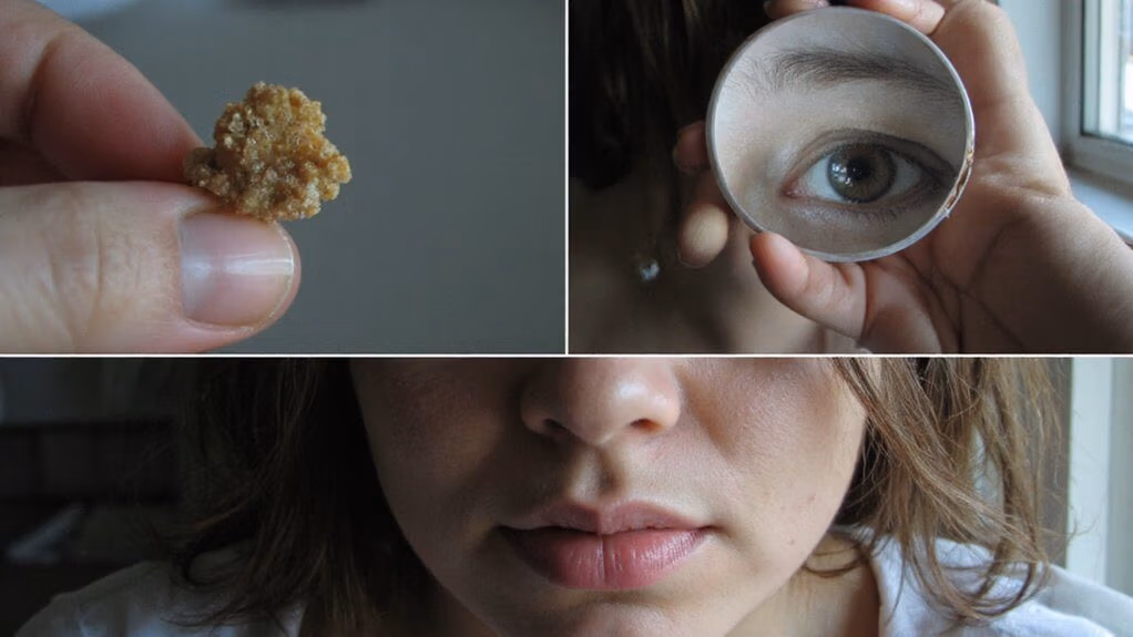
Because iron is essential for erythropoiesis and for iron-dependent enzymes that support keratinocyte and follicular proliferation, deficiency commonly produces characteristic cutaneous, hair, and nail findings. You’ll notice pallor of the conjunctiva and generalized skin hypopigmentation due to reduced hemoglobin and altered melanogenesis. Hair becomes brittle, with increased telogen effluvium frequency and slowed anagen recovery; follicular miniaturization can occur in chronic deficiency. Nails show koilonychia, brittle ridging, and longitudinal melanonychia in some cases. You should evaluate iron indices when these findings are unexplained.
- Conjunctival and cutaneous pallor correlating with ferritin and hemoglobin levels
- Telogen effluvium with delayed anagen re-entry
- Koilonychia and structural nail plate abnormalities
- Keratinocyte proliferation impairment on biopsy
You should integrate dermoscopy and targeted biopsy to quantify pathology and guide novel therapeutic strategies.
Restless Legs and Sleep Disturbances
You should be aware that iron deficiency is associated with increased risk of restless legs syndrome (RLS) via reduced brain iron and impaired dopaminergic function. This often manifests as nocturnal leg urges and dysesthesias that provoke movements, increasing sleep fragmentation and lowering sleep efficiency. You should assess ferritin and transferrin saturation when patients report nighttime leg urges or fragmented sleep to guide targeted iron repletion.
Restless Legs Connection
Although classically considered a hematologic disorder, iron deficiency contributes directly to restless legs syndrome (RLS) and associated sleep fragmentation by reducing central nervous system iron stores and impairing dopamine synthesis.
- Target ferritin thresholds for symptomatic RLS evaluation (often <50 ng/mL).
- Use transferrin saturation and CRP to interpret ferritin.
- Consider neuroimaging or CSF studies for refractory cases.
- Prefer IV iron when oral absorption is inadequate or rapid repletion is needed.
You should evaluate nocturnal sensory-motor discomfort with focused history and neurological exam, and you’ll monitor objective symptom change after repletion. Emerging strategies target iron transport and neurochemical coupling to innovate therapeutics. You should integrate multidisciplinary care, incorporate patient-reported outcome measures, and collaborate with researchers to implement translational diagnostics and adaptive treatment algorithms for precision medicine.
Sleep Fragmentation Risk
When RLS-related sensorimotor symptoms occur nocturnally, they fragment sleep architecture by provoking periodic limb movements (PLMS), transient cortical arousals, and reductions in sleep efficiency and slow-wave sleep. You’ll experience increased sleep fragmentation that elevates nocturnal sympathetic tone, impairs memory consolidation, and degrades restorative processes mediated by slow-wave activity. Objective polysomnography often shows higher arousal indices, decreased REM continuity, and microarousal clustering temporally linked to PLMS bursts. Iron deficiency exacerbates dopaminergic and adenosinergic dysfunction implicated in PLMS pathophysiology, so correcting iron status can normalize PLMS frequency and improve macro- and microarchitecture metrics. Clinically, you should assess ferritin and transferrin saturation when sleep fragmentation is prominent, integrate actigraphy or PSG for quantification, and consider evidence-based iron repletion to restore sleep integrity while monitoring therapeutic response closely.
Nighttime Leg Urges
If you experience nocturnal sensory-motor urges characteristic of restless legs syndrome (RLS), they’ll present as an uncomfortable, often dysesthetic need to move the legs that emerges or worsens at rest and in the evening, disrupting sleep initiation and maintenance. You should evaluate for iron deficiency because low central and peripheral iron correlates with RLS severity; ferritin <50 µg/L often associates with symptoms. Assess ferritin, transferrin saturation, and inflammatory markers, and consider dopaminergic and α2δ ligand responsiveness for symptomatic profiling. Treating iron deficiency can reduce periodic limb movements and sleep fragmentation. Innovations in chelatable iron imaging and targeted supplementation protocols are promising.
- Ferritin threshold and symptom correlation
- Transferrin saturation and diagnostic yield
- Response to iron repletion vs pharmacotherapy
- Emerging neuroimaging biomarkers
Implement protocolized monitoring and follow-up.
Digestive Symptoms and Appetite Changes
Because iron is integral to mucosal integrity, neurotransmitter synthesis, and epithelial cell function, iron deficiency often produces measurable gastrointestinal signs and alters appetite. You may notice dysphagia, glossitis-associated taste change, or atrophic gastritis symptoms; these correlate with impaired mucosal repair and altered serotonin/dopamine signalling affecting satiety and craving. You might develop pica or specific nonfood cravings, reduced hunger, or early satiety related to delayed gastric emptying. Objective assessment includes gastrointestinal history, hemoglobin, ferritin, transferrin saturation, and targeted endoscopy when indicated. Interpret results with protocols that prioritize biomarker variability and patient-reported appetite shifts. You should integrate therapeutics, iron repletion strategy, and motility-targeted interventions per guideline consensus.
| Symptom | Mechanism | Clinical action |
|---|---|---|
| Pica/craving | Neurotransmitter dysregulation | Assess ferritin |
| Early satiety | Motility alteration | Gastric emptying studies |
| Dysgeusia | Mucosal atrophy | Oral exam and biopsy |
How Iron Deficiency Shows in Children and Teens
In children and adolescents, iron deficiency commonly presents with neurodevelopmental, behavioral, and growth-related findings rather than the classic adult symptom complex. You may observe cognitive slowing, attention deficits, and impaired executive function that reduce academic performance; these correlate with brain iron-dependent neurotransmitter synthesis. You may notice irritability, apathy, or pica behaviors reflecting altered reward circuits. Growth retardation and delayed skeletal maturation can occur due to impaired oxygen delivery and metabolic dysregulation. Laboratory-confirmed iron deficiency also associates with restless legs–type movements and altered sleep architecture. Clinically assess symptom clusters and trajectory rather than isolated signs. Integrate developmental surveillance with objective measures.
- Cognitive and attentional decline impacting learning
- Behavioral changes including pica and irritability
- Linear growth deceleration and delayed bone age
- Sleep disturbances and hyperkinetic movements
When to Seek Medical Testing
When you or your child has persistent symptoms suggestive of iron deficiency or belongs to a recognized high‑risk group, initiate guideline‑directed laboratory evaluation without delay. You should request CBC with indices, ferritin, transferrin saturation, and CRP to contextualize inflammatory effects on ferritin. Prioritize age‑specific reference ranges and repeat testing when initial results are equivocal. Consider targeted investigations for blood loss, malabsorption, or chronic disease based on history and exam.
| Indication | Action |
|---|---|
| Microcytic anemia | Order ferritin, TSAT |
| Low ferritin | Evaluate sources of iron loss |
| High CRP with normal ferritin | Reassess with soluble transferrin receptor |
| High-risk patient | Screen per guideline frequency |
Document results, communicate urgency, and arrange specialist referral when diagnostic uncertainty or severe anemia exists. You should use validated algorithms and update care plans rapidly.
Treatment Approaches and Follow-Up
Although oral iron remains first‑line for most patients, you should individualize treatment by severity, rate of hemoglobin decline, absorption capacity, and tolerance. You’ll select oral ferrous salts or ferric maltol when GI tolerance and absorption are adequate; dose to replenish iron deficit (typically three to six mg/kg elemental iron per day in pediatrics or 100–200 mg elemental iron per day in adults) and continue three months after hemoglobin normalization. Consider IV iron for malabsorption, ongoing losses, intolerance, or rapid repletion needs. Monitor CBC and ferritin at 4–8 weeks and adjust therapy.
- Use IV formulations based on risk profile and infusion time.
- Employ pharmacogenomic or microbiome considerations selectively.
- Integrate adherence technology and remote monitoring.
- Escalate to transfusion only for hemodynamic instability and personalized risk stratification.
Conclusion
You should recognize that mucocutaneous changes (pallor, glossitis, angular cheilosis, koilonychia), neurocognitive symptoms, dyspnea, tachycardia, and gastrointestinal alterations commonly reflect iron deficiency. Correlate clinical signs with CBC, serum ferritin, transferrin saturation and CRP to distinguish true iron deficiency from inflammatory sequestration. If abnormalities persist, pursue etiologic evaluation (GI blood loss, malabsorption, dietary insufficiency) and institute iron repletion with monitoring of hematologic and iron indices. Adjust therapy by response and adverse effects; refer promptly when indicated.
