You may have a peptic (stomach) ulcer if you notice a persistent burning or gnawing pain in your upper abdomen that often worsens between meals or at night. You’ll commonly get dyspepsia, bloating, nausea, reduced appetite, or unexplained weight loss; vomiting blood or black stools are alarm signs—knowing which symptoms need urgent evaluation will shape what you do next.
Common Symptoms and Early Warning Signs
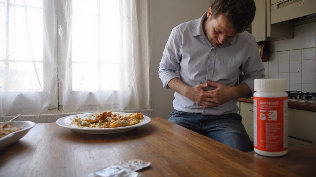
Often you’ll notice a gnawing or burning pain in the upper abdomen that comes and goes, frequently worsening between meals or at night and often temporarily relieved by antacids. You may also experience persistent dyspepsia, nausea, bloating, early satiety and reduced appetite. Recurrent vomiting, black or tarry stools, or visible blood indicate mucosal breach and require urgent evaluation. Chronic bleeding can produce iron-deficiency anemia—fatigue, pallor, dyspnea on exertion—and laboratory testing will detect low hemoglobin and ferritin. Unintentional weight loss and failure to respond to standard gastroesophageal treatments raise suspicion for complicated ulcer disease or malignancy. Risk stratification includes NSAID use, smoking and confirmed Helicobacter pylori infection; targeted testing and endoscopic assessment guide management. Act promptly to optimize diagnostic yield and therapeutic innovation and outcomes.
Pain Patterns and Timing
You may feel a burning or gnawing pain in the upper abdomen, typically between the breastbone and navel. Pain timing varies with meals—some ulcers worsen soon after eating while others briefly improve with food and recur hours later. Nighttime or early-morning pain that wakes you or follows prolonged fasting is common and warrants clinical evaluation.
Location of Pain
Where pain is felt gives important diagnostic clues: peptic ulcers typically produce a midline epigastric ache or burning sensation that can radiate to the back or to the right or left upper quadrant. You should note exact location, lateralization, and any radiation pattern, as these guide differential diagnosis and imaging choices. Document severity, relation to posture, and concomitant signs (nausea, vomiting, weight loss). Use objective mapping to track progression and response to therapy.
- Midline epigastric burning suggests gastric or duodenal ulcer
- Radiation to the back raises concern for posterior penetration
- Right upper quadrant pain may mimic biliary disease
- Left upper quadrant radiation is less common but possible
- Diffuse or shifting pain prompts broader evaluation
Correlate findings with endoscopy and targeted imaging expediently as indicated.
Pain Timing With Meals
Because meal timing reliably differentiates ulcer types, document whether pain improves with food, appears within 30–60 minutes of eating, or recurs several hours later; duodenal ulcers classically produce hunger-like epigastric pain that’s relieved by eating then returns 2–4 hours later or at night, while gastric ulcers more commonly cause immediate postprandial pain and anorexia with weight loss. You should assess pattern: ask how soon pain starts after meals, which foods modify symptoms, and whether antacids or meals relieve pain. Correlate timing with risk factors—NSAID use, H. pylori exposure, smoking—to prioritize diagnostics. Use timing to guide urgent testing vs outpatient evaluation: postprandial pain with weight loss warrants prompt endoscopic assessment; meal-relieved, cyclic pain suggests trial of eradication and acid suppression with follow-up and careful monitoring.
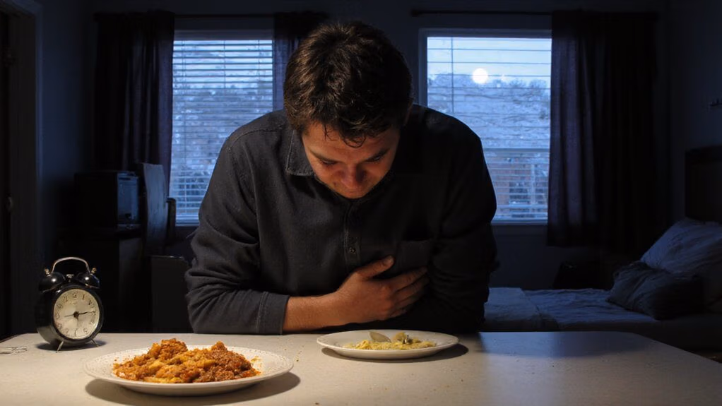
Nighttime and Early-Morning Pain
Often patients report epigastric pain that wakes them from sleep or occurs in the early morning hours, a pattern classically associated with duodenal ulcer disease. You should note timing because nocturnal pain suggests acid-mediated pathology and may guide testing and treatment. You’ll be asked about pain onset, duration, relation to food, and response to antacids. Objective assessment can include H. pylori testing and endoscopy when alarm features exist. Consider risk stratification to prioritize diagnostics and novel point-of-care breath tests when available.
- Pain awakening from sleep often reflects increased nocturnal acid secretion
- Early-morning discomfort may improve with food or antacid
- Nocturnal pain increases likelihood of duodenal ulcer
- Alarm signs necessitate urgent evaluation
- Use targeted testing to confirm diagnosis
Document patterns and reassess response to therapy.
Heartburn and Acid Reflux

You may experience a burning chest sensation, regurgitation, sour taste, or throat irritation—symptoms consistent with gastroesophageal reflux. These symptoms often worsen after meals, when lying flat, or with bending. Common triggers include fatty or spicy foods, citrus, caffeine, alcohol, and smoking, and identifying them helps differentiate reflux from ulcer-related pain.
Typical Heartburn Symptoms
How does heartburn present clinically? You typically report a retrosternal burning sensation that rises toward the throat, often accompanied by regurgitation of sour fluid. Symptoms are intermittent, relate to meals or posture, and may mimic cardiac pain; you should distinguish based on quality and timing. Objective testing (endoscopy, pH monitoring) clarifies diagnosis when needed.
- Retrosternal burning or warmth
- Acid regurgitation or sour taste
- Hypersalivation or unpleasant throat sensation
- Nocturnal symptom worsening disrupting sleep
- Episodic onset with variable intensity
You should seek evaluation if symptoms are severe, persistent, or atypical; evidence-based assessment guides targeted therapy and monitoring. Early intervention reduces complications and tailors advanced management options such as pharmacologic acid suppression, endoscopic assessment, or surgical referral when evidence indicates. Monitor response and adjust care promptly.
Common Acid Reflux Triggers
Because foods, behaviors, and medications that lower lower‑esophageal sphincter tone or raise gastric acidity will precipitate reflux, you’ll commonly see predictable triggers across patients: high‑fat meals, chocolate, caffeine, alcohol, spicy or acidic foods (citrus, tomato), and carbonated beverages; large or late meals and recumbency after eating promote reflux by increasing gastric distention and pressure; additionally, obesity, smoking, pregnancy, NSAIDs, and medications that relax the LES (eg, nitrates, calcium‑channel blockers, certain anticholinergics) increase symptom frequency and severity. Assess trigger exposure and temporal links to symptoms, then prioritize targeted modifications: reduce late or large meals, limit alcohol and caffeine, avoid trigger foods, and stop smoking. Consider weight reduction and medication review to replace LES‑relaxing agents when feasible. Use objective monitoring and iterative adjustments to optimize control.
Nausea, Vomiting, and Blood in Vomit
When a peptic ulcer erodes a mucosal vessel, you can develop nausea and vomiting that may contain blood (hematemesis), which often appears as bright red or “coffee-ground” material depending on bleeding rate and gastric digestion. You should treat hematemesis as a potential emergency: it signals mucosal disruption with risk of hemodynamic compromise. You’ll also notice premonitory nausea, retching, and variable pain. Diagnostics include urgent assessment of vital signs, hemoglobin, coagulation studies, and endoscopic evaluation to localize and control bleeding. Management integrates resuscitation, proton pump inhibition, endoscopic hemostasis, and targeted therapy for underlying causes.
- Recognize bright red versus coffee-ground emesis
- Monitor for hypotension and tachycardia
- Obtain lab tests promptly
- Arrange urgent endoscopy
- Initiate acid suppression therapy
Follow local protocols and guidelines.
Changes in Appetite and Unexplained Weight Loss
After episodes of nausea, vomiting, or bleeding you may notice decreased appetite and unintended weight loss; these changes often reflect ongoing pain with eating, food avoidance from fear of symptoms, or more advanced disease processes. You should track weight, meal patterns, and symptom timing to quantify decline and guide evaluation. Ulcer-related anorexia and cachexia correlate with persistent mucosal inflammation, impaired gastric function, or complications like gastric outlet obstruction or malignancy; endoscopic assessment and H. pylori testing are evidence-based next steps. Nutritional deficits can worsen outcomes, so initiate targeted interventions—dietitian referral, caloric supplementation, and treat the underlying ulcer promptly. If weight loss is rapid (>5% in one month) or accompanied by progressive dysphagia or anemia, prioritize urgent specialist referral and diagnostic imaging to expedite care delivery.
Bloating, Belching, and Indigestion
How often do bloating, belching, and indigestion point to an ulcer? You may notice persistent postprandial fullness, repeated belching, and dyspepsia that don’t respond to routine measures; these symptoms can reflect mucosal irritation or delayed gastric emptying associated with peptic ulcer disease. Evaluate symptom timing, relation to meals, and response to antacids; if symptoms are recurrent or progressive, pursue diagnostic testing such as endoscopy or noninvasive H. pylori testing. Use targeted, data-driven interventions rather than empiric escalation. Monitor for alarm features (weight loss, anemia, persistent vomiting).
- Persistent post-meal fullness
- Recurrent, unexplained belching
- Epigastric burning or discomfort
- Poor response to standard antacids
- Symptoms worsening over weeks
Act promptly to confirm diagnosis and guide therapy. Integrate novel diagnostics and personalized treatment pathways where evidence supports benefit.
Stool Changes and Black or Tarry Stools
Why is a stool black or tarry, and what does that tell you clinically? Black, tarry stool (melena) signals upper gastrointestinal bleeding; hemoglobin is digested and darkens stool. You should note onset, amount, and associated symptoms (dizziness, tachycardia). Immediate evaluation—nasogastric aspirate, hemoglobin, endoscopy—guides diagnosis and therapy. Mild darkening from iron or bismuth differs; medication history matters. Track stool changes and report melena promptly; prompt endoscopic visualization reduces morbidity. Below is a concise visual guide.
| Finding | Clinical implication |
|---|---|
| Melena | Upper GI bleed; urgent endoscopy |
| Black from iron/bismuth | Medication-related; not bleeding |
| Occult blood positive | Occult bleeding; evaluate labs |
| Bright red blood per rectum | Lower GI or brisk upper bleed; alternate workup |
If you see these changes, seek urgent medical assessment; coordinated care and endoscopy improve outcomes.
Risk Factors That Intensify Symptoms
Recognize that certain medications, infections, and comorbid conditions markedly worsen ulcer symptoms and bleeding risk. You should note that NSAIDs impair mucosal defense, while anticoagulants increase hemorrhage potential; H. pylori infection provokes persistent inflammation and delays healing. Chronic diseases like liver cirrhosis or chronic kidney disease amplify vulnerability, and smoking or high-dose corticosteroids further impair repair mechanisms. Assess combined risk quantitatively to guide therapy selection and monitoring.
- NSAIDs and aspirin
- Anticoagulants (warfarin, DOACs)
- H. pylori infection
- Chronic liver or renal disease
- Smoking and high-dose corticosteroids
You should prioritize testing for H. pylori, review medication lists for deprescribing opportunities, consider gastroprotection with proton pump inhibitors when indicated, and implement smoking cessation and multidisciplinary care pathways to reduce symptom severity and bleeding complications in high-risk patients.
When to Seek Immediate Medical Care
If you have sudden, severe upper abdominal pain, vomit blood (hematemesis), pass black or tarry stools (melena), or become lightheaded or faint, seek emergency care immediately. You should also get urgent evaluation if pain is progressive despite medications, you develop persistent vomiting, unexplained weight loss, or signs of infection such as fever and tachycardia. Notify clinicians about NSAID use, anticoagulants, or prior peptic ulcer disease; these raise bleeding risk and change management. Emergency assessment will include airway, breathing, circulation stabilization, IV access, labs, and urgent endoscopy when indicated. Early intervention reduces morbidity and mortality from hemorrhage or perforation. If you’re using digital health tools, confirm real-time sharing of physiologic data and medications with responders to accelerate care. Follow clinician instructions for diagnostics and definitive therapy.
Conclusion
If you have persistent epigastric pain, dyspepsia, unexplained weight loss, vomiting, hematemesis, melena, or iron‑deficiency anemia, seek evaluation promptly. Risk factors like NSAID use and H. pylori increase likelihood of peptic ulcer disease; you’ll need testing (noninvasive H. pylori or endoscopy) and targeted treatment. Don’t ignore alarm features or progressive symptoms — timely diagnosis and evidence‑based management reduce complications such as bleeding and perforation. Follow-up care and eradication therapy markedly improve outcomes when adhered to consistently.